跳转到内容

File:Surface projections of the organs of the trunk.png

页面内容不支持其他语言。
這個文件來自維基共享資源
维基百科,自由的百科全书

原始文件 (1,583 × 2,533像素,文件大小:3.33 MB,MIME类型:image/png


摘要

描述
English: Surface projections of the major organs of the trunk, using the vertebral column and rib cage as main reference points of superficial anatomy. The transpyloric plane and McBurney's point are among the marked locations.

To discuss image, please see Talk:Human body diagrams

日期
来源

All included images are in public domain. See Template:Human body diagrams for individual details.

References

Spinal vertebrae levels:

  • Located at middle of frontal face of spinal vertebrae according to reference the image :File:Gray 111 - Vertebral column.png when pasted into image to superiorly fit the surface bulges of the clavicular bones and inferiorly fit the surface bulges of the anterior iliac spine.

Gross overview of organ locations:

  • Plate 260B - Regions and Planes of Adbomen. From: The Interactive Atlas of Human Anatomy Version 3.0 (CD-ROM). Authors: Frank H. Netter and Carlos A. G. Machado. Published by Saunders, October 1, 2002. ISBN-10: 1929007140. ISBN-13: 978-1929007141.
  • Fig 139 from The SPENCER SYSTEM. SEVENTEENTH EDITION
  • Plate 1034 from Gray's Anatomy

Locations of specific organs:
The following vertebral levels are generally given by the middle of the vertebral body.

  • Bålens ytanatomy (Superficial anatomy of the trunk). Anca Dragomir, Mats Hjortberg and Godfried M. Romans. Section for human anatomy at the Department of medical cell biology, Uppsala university, Sweden.
Key points from that compendium:
  • The oblique fissure of the right lung goes from the intercostal space 4 to 6
  • The horizontal fissure goes along the 4th rib (the original image from Gray has this line almost at the 5th rib, but this is not labeled in image, as other lung images seem to support the upper level)
  • The cardia of the stomach is at the level of the 10th rib
  • The fundus of the stomach is in the 5th intercostal space, slightly below the apex of the heart
  • Pylorus is usually at the L1 (the transpyloric plane)
  • The transpyloric plane is at the level of:
    • lumbar vertebra 1
    • the pancreatic body
    • the origins of the superior mesenteric artery from the aorta and portal vein
    • the left and right colic flexure
    • hilum of the kidney on the left
    • upper pole of the kidney on the right
    • duodenojejunal flexure
  • McBurney's point is located one third of a line from the anterior superior iliac spine and the navel
  • The upper rim of the liver is in the 5th intercostal space
  • The left lobe of the liver is to the left of the midline
  • The lower limit of the liver at the midline is approximately 3 cm below processus xiphoideus
  • The spleen is projected against the 9th and 10th rib on the backside
  • The left kidney is approximately at the vertebral level T12 to L3, and the right one slightly lower.
  • Atlas of Human Anatomy in Cross Section: Appendix: Topography of the Thorax and Abdomen. Ronald A. Bergman, Ph.D., Adel K. Afifi, M.D., Jean J. Jew, M.D., and Paul C. Reimann, B.S. [1]:
  • The average position of the highest point of the spleen is at the level of the lower third of the tenth thoracic vertebra.
  • The average position of the lowest point of the spleen is at the level of the middle third of the first lumbar vertebra.

Position of the navel

Urinary tract:

作者 Mikael Häggström
File:Surface projections of the organs of the trunk.svg是此文件的矢量版本。 如果此文件质量不低于原点阵图,就应该将这个PNG格式文件替换为此文件。

File:Surface projections of the organs of the trunk.png → File:Surface projections of the organs of the trunk.svg

更多信息请参阅Help:SVG/zh

其他语言
Alemannisch  العربية  беларуская (тарашкевіца)  български  বাংলা  català  нохчийн  čeština  dansk  Deutsch  Ελληνικά  English  British English  Esperanto  español  eesti  euskara  فارسی  suomi  français  Frysk  galego  Alemannisch  עברית  hrvatski  magyar  հայերեն  Bahasa Indonesia  Ido  italiano  日本語  ქართული  한국어  lietuvių  македонски  മലയാളം  Bahasa Melayu  norsk bokmål  Plattdüütsch  Nederlands  norsk nynorsk  norsk  occitan  polski  prūsiskan  português  português do Brasil  română  русский  sicilianu  Scots  slovenčina  slovenščina  српски / srpski  svenska  தமிழ்  ไทย  Türkçe  татарча / tatarça  українська  vèneto  Tiếng Việt  中文  中文(中国大陆)  中文(简体)  中文(繁體)  中文(马来西亚)  中文(新加坡)  中文(臺灣)  中文(臺灣)  +/−
新SVG图片

许可协议

Public domain 我,本作品著作权人,释出本作品至公有领域。这适用于全世界。
在一些国家这可能不合法;如果是这样的话,那么:
我无条件地授予任何人以任何目的使用本作品的权利,除非这些条件是法律规定所必需的。

Human body diagrams

Main article at: Human body diagrams

Template location:Template:Human body diagrams

How to derive an image

Derive directly from raster image with organs

The raster (.png format) images below have most commonly used organs already included, and text and lines can be added in almost any graphics editor. This is the easiest method, but does not leave any room for customizing what organs are shown.

Adding text and lines:

Derive "from scratch"

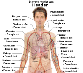
By this method, body diagrams can be derived by pasting organs into one of the "plain" body images shown below. This method requires a graphics editor that can handle transparent images, in order to avoid white squares around the organs when pasting onto the body image. Pictures of organs are found on the project's main page. These were originally adapted to fit the male shadow/silhouette.

Organs:

More organs are found at: Human body diagrams/Organs

Derive by vector template

The Vector templates below can be used to derive images with, for example, Inkscape. This is the method with the greatest potential.

See Human body diagrams/Inkscape tutorial for a basic description in how to do this.

Examples of derived works

More examples

Licensing

Creative Commons CC-Zero 本作品采用知识共享CC0 1.0 通用公有领域贡献许可协议授权。
采用本宣告发表本作品的人,已在法律允许的范围内,通过在全世界放弃其对本作品拥有的著作权法规定的所有权利(包括所有相关权利),将本作品贡献至公有领域。您可以复制、修改、传播和表演本作品,将其用于商业目的,无需要求授权。

说明

添加一行文字以描述该文件所表现的内容

此文件中描述的项目

描繪內容

文件历史

点击某个日期/时间查看对应时刻的文件。

最新 | 最旧) 查看(较新10条 | )(10 | 20 | 50 | 100 | 250 | 500
日期/时间缩⁠略⁠图大小用户备注
当前2019年12月27日 (五) 09:192019年12月27日 (五) 09:19版本的缩略图1,583 × 2,533(3.33 MB)Mikael Häggström+Costal margin
2010年11月11日 (四) 10:382010年11月11日 (四) 10:38版本的缩略图1,050 × 1,680(2.07 MB)Mikael HäggströmAdapted to recently added overview images. Distinguished different ways to designate vertebrae levels.
2010年11月7日 (日) 10:042010年11月7日 (日) 10:04版本的缩略图936 × 1,325(1.77 MB)Mikael Häggströmupdate from svg
2010年11月7日 (日) 09:462010年11月7日 (日) 09:46版本的缩略图936 × 1,325(1.77 MB)Mikael Häggströmupdate from svg
2010年10月24日 (日) 04:512010年10月24日 (日) 04:51版本的缩略图936 × 1,325(1.61 MB)Mikael HäggströmSmoother edges
2010年10月10日 (日) 05:182010年10月10日 (日) 05:18版本的缩略图936 × 1,325(1.61 MB)Mikael HäggströmMinor kidney adjustment. More realistic hip bone
2010年10月6日 (三) 04:472010年10月6日 (三) 04:47版本的缩略图936 × 1,325(1.73 MB)Mikael HäggströmDistinguished stomach and spleen. Removed painted arteries out of scope.
2010年10月4日 (一) 18:402010年10月4日 (一) 18:40版本的缩略图936 × 1,325(1.74 MB)Mikael HäggströmLowered spleen
2010年10月3日 (日) 15:212010年10月3日 (日) 15:21版本的缩略图936 × 1,325(1.74 MB)Mikael HäggströmDecreased some opacity. Aligned tail of pancreas with spleen. Adjusted fissure marking width.
2010年10月2日 (六) 18:202010年10月2日 (六) 18:20版本的缩略图936 × 1,325(1.72 MB)Mikael Häggström+liver label
最新 | 最旧) 查看(较新10条 | )(10 | 20 | 50 | 100 | 250 | 500

以下3个页面使用本文件:

全域文件用途

元数据